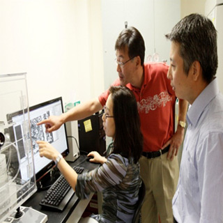
Currently available implant design is probably unable to understand bacterial infection, a major cause of failure in orthopedic implants. Apparently developing infection-fighting drugs or biomaterials is a task because of insufficient laboratory equipment. During the research, scientists seeded 0.02 mL microfluidic channels with osteoblasts and immunized the channels with Staphylococcus epidermis bacteria. This bacterium is known as a common pathogen in orthopedic infections. Experts pumped nutrient solutions by means of channels at a concentration and flow rate mimicking conditions within the human body.
Then with the help of a microscope, bone tissue cells and bacteria within the channels were imaged. These images were inspected for bacteria count. Dr. Joung-Hyun Lee of Stevens, of the New Jersey Dental School and colleagues observed that microfluidic devices and finely-tuned dynamic flow settings show realistic bone tissue models in clinical scenarios. In this system microfluidic channels seemingly demonstrate a realistic environment for cells to grow and stick to three dimensions. In addition, dynamic fluid motion through the channels apparently imitates real-world conditions, which were previously unattainable in a lab setting. This fluid was probably equipped with solutions potentially carrying antibiotics or other unique drugs.
The research is published in the journal Tissue Engineering.
